Ferns are common plants all over the world. They are often been neglected and unappreciated, which may be due to their lack of colorful flowers and the fact that they only have different shades of green. Yet, their presence occupies spaces where other flowering plants are not suitable to grow. Also, their horizontal “stems” and shallowed “roots” can help preventing soil erosion.
In Taxonomy, ferns are classified as vascular plants since they have vascular systems, which transport water and nutrients efficiently. Unlike other vascular plants such as the flowering plants (angiosperms) and conifers (gymnosperms), ferns reproduce via spores, while angiosperms and gymnosperms reproduce via seeds.
There are 3 major parts of ferns that we can observe:
The fronds, being responsible for carrying out photosynthesis, might be dimorphic that one is the sterile frond (cannot produce spores) and the other one is fertile frond (reproductive, can produce spores) ;
The rhizome;
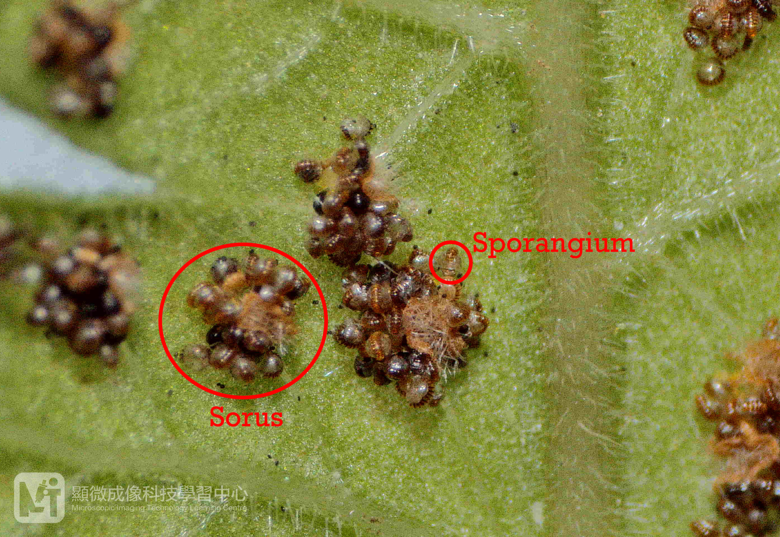
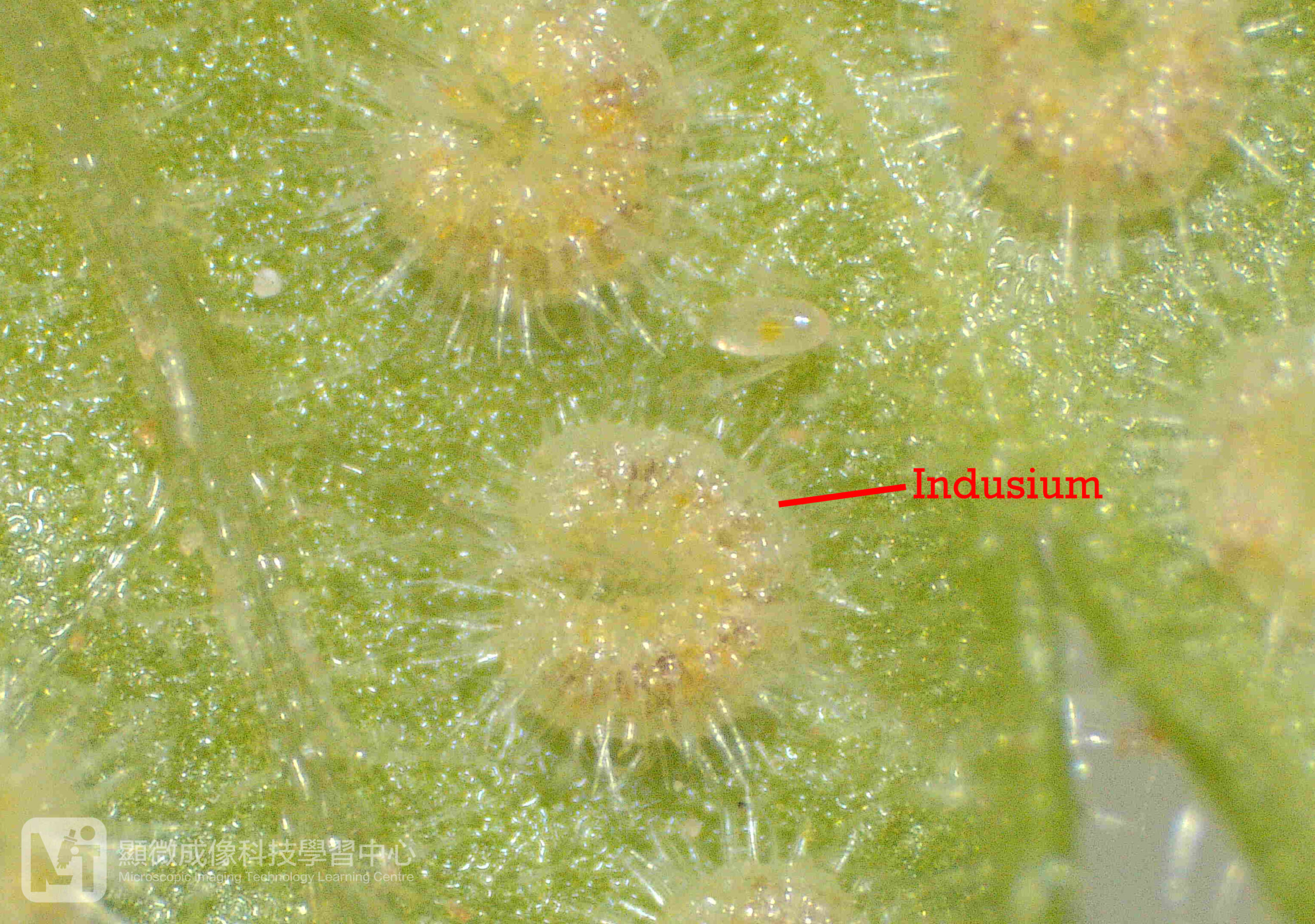
The reproductive structures called sporangia. The sporangia are found on the lower surface of the fertile frond. The clusters of sporangia are known as sori. Sori may be exposed or protected by a cover called the indusium.
We can identify different types of ferns not only by the appearance of their fronds, but also the position and distribution of the sporangia at the lower surface of the fronds, the shape of the indusium and the sporangia, as well as the appearance of the spores. However, the sporangia and spores are very tiny, different kinds of microscopes are used to observe them.
1. Observing sporangia under a stereomicroscope
It is easy to observe the specimens by using a stereomicroscope. Simply put the fern frond specimens on the stage of the stereomicroscope and use different magnifications to observe them. The range of magnification is around 6x to 50x. We can get a 3D colorful image under the stereomicroscope.
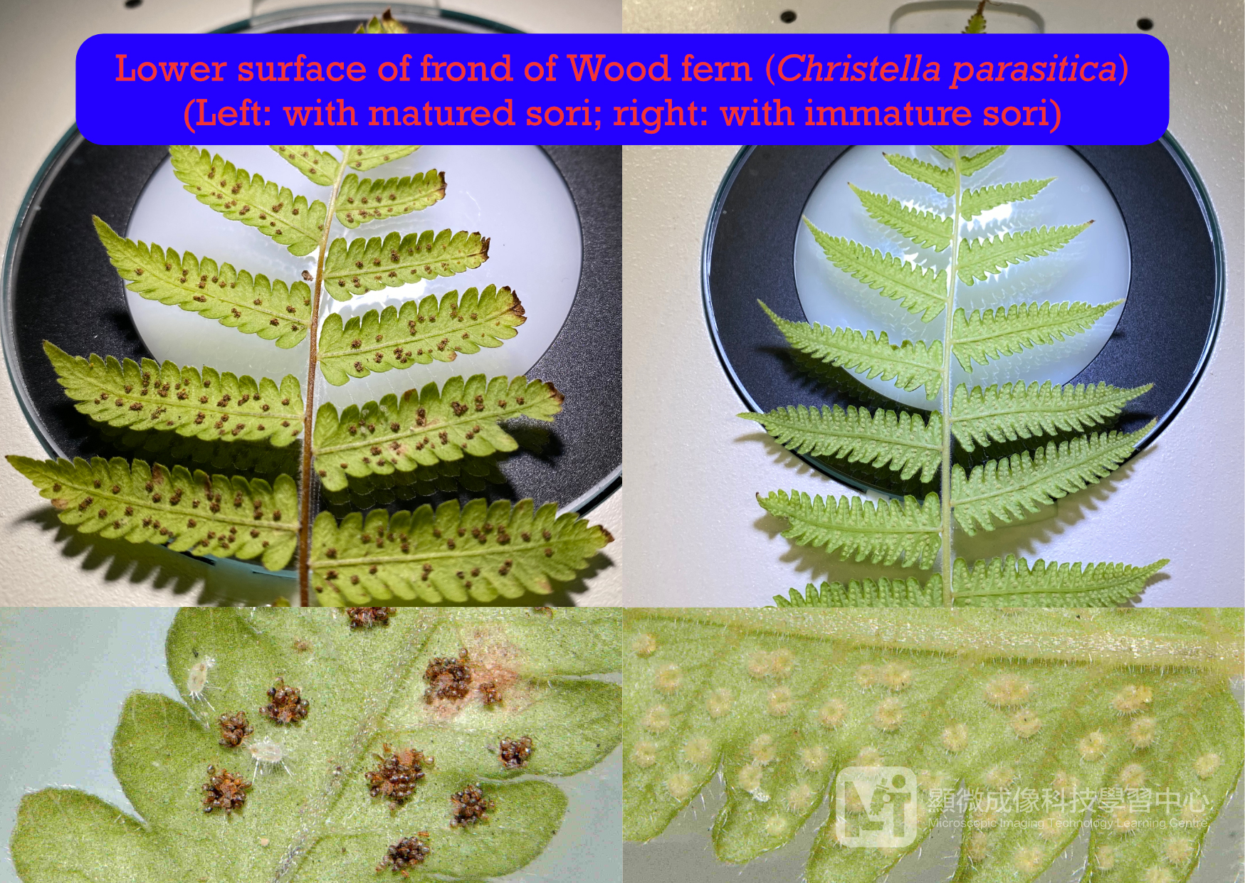
Figure 1. Lower surface of frond of Wood fern (Christella parasitica)Christella parasitica ) 的葉底
Figure 2. Sori and Sporangia of Wood fern (Christella parasitica) (x30)
Figure 3. Indusium on the sorus of Wood fern (Christella parasitica) (x50)
2. Observing sporangia and spores under a compound microscope
Before observing the sporangia and spore under compound microscope, we must first prepare the slides. How to prepare the slides? We can follow the steps as shown below:
1. 1. Add a drop of water on the slide;
2. 2. Use a pair of forceps to transfer the sporangia and spores into the water;
3. 3. Cover the specimens with a cover slip.
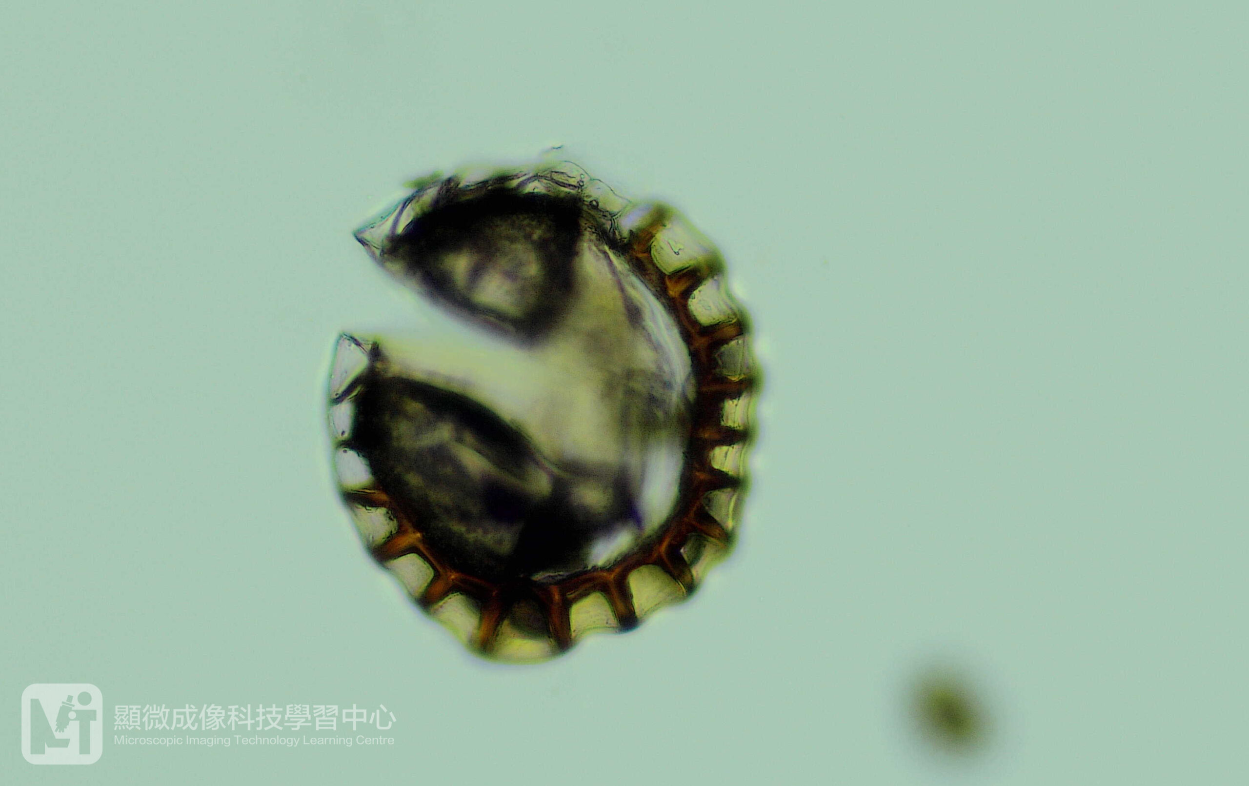
Now the specimen is ready to observe. The range of magnification is 40x to 400x. We can get a 2-D colorful image under compound microscopes.
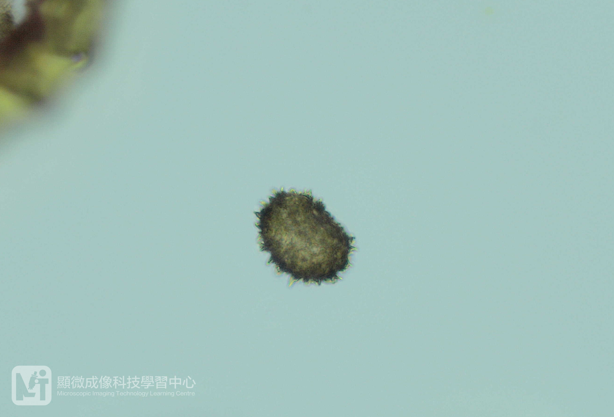
Figure 4. A sporangium of Wood fern (Christella parasitica) (x200)
Figure 5. A spore of Wood fern (Christella parasitica) (x400)
3. Observing the sporangia and spores under a scanning electron microscope (SEM)
For observing the sporangia and spores under a SEM, we should put the specimens on a metal stage with a conductive adhesive tape stuck on it. Place the stage and the specimens into a sputter coater for coating a thin layer of metal on the surface of the specimens. Then it is ready to observe under the SEM. The range of magnification is 60x to 30,000x. The image generated from the SEM is a 3D and black-and-white image showing the exterior surface of a specimen.
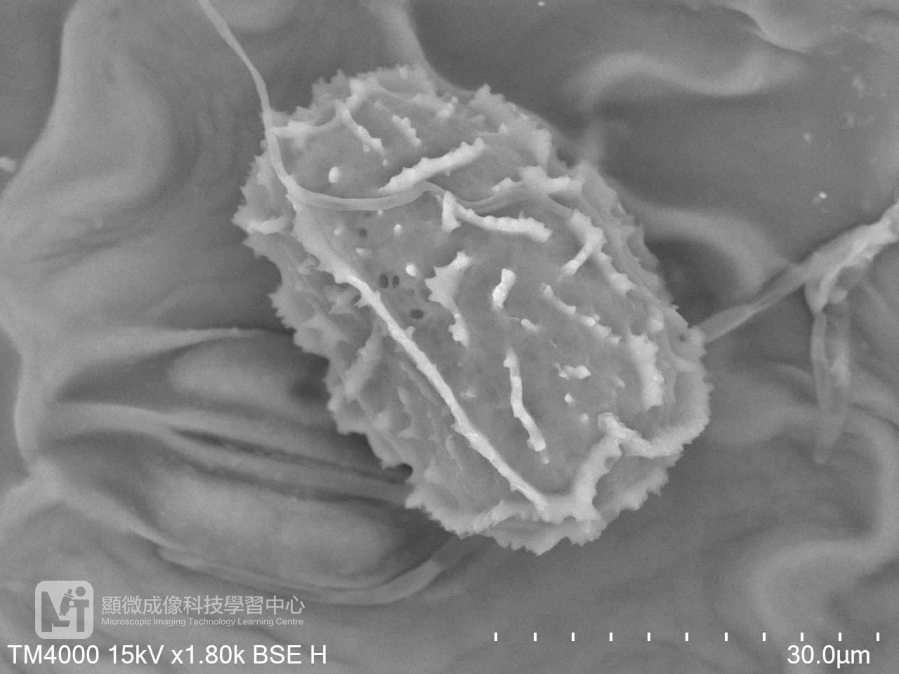
Figure 6. A spore of Wood fern (Christella parasitica) (x1,800)
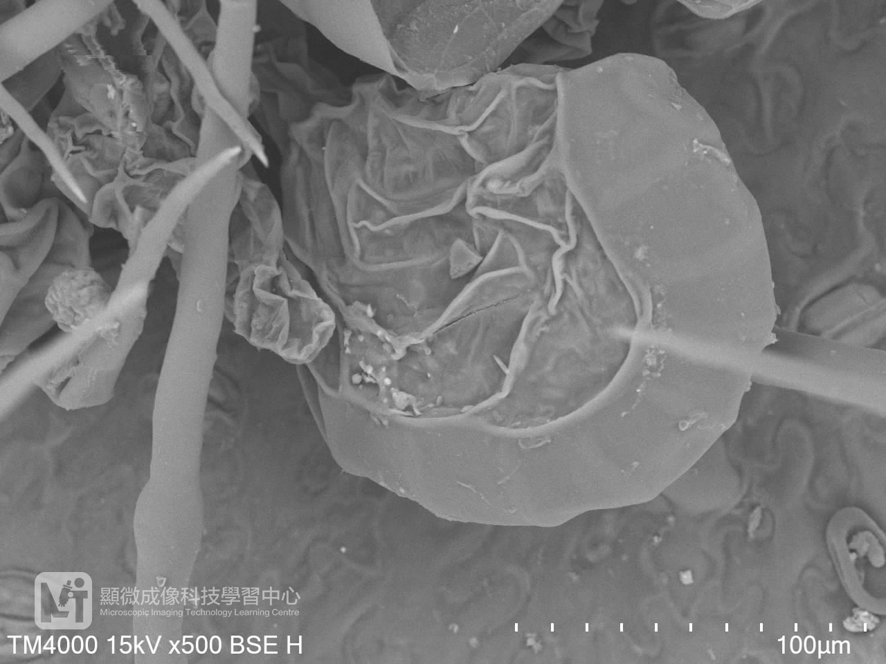
Figure 7. A sporangium of Wood fern (Christella parasitica) (x500)
As the external features of sporangia and spore of different fern species varies, microscopes are very useful tools for us to identify them. Different kinds of microscopes allow us to observe distinctive details of them. Ferns are very interesting plants, you might try to explore more about them!