There has been much debate about who the inventor of microscopes is. Generally, historians believes that the first microscope was invented by Hans and Zacharias Janssen, the Dutch spectacles makers, in the 1590s. It is a microscope consisting of a tripod, a main tube with 1-2 inches of diameter, a base disk, a concave lens and a convex lens (American Physical Society, 2004). With only 3x to 9x magnification and rough image quality (American Physical Society, 2004), the microscope was not widely used by scientists.
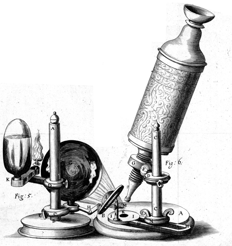
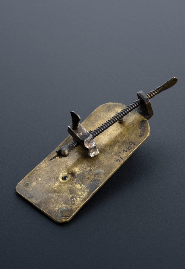
Until the 1660s, a natural philosopher (an early scientist) Robert Hooke improved the design of the microscope including enhancing the lens and illumination systems. However, the image acquired was still blurred (Davidson, 2010). Besides, the focusing system was difficult to use and it easily caused uneven wearing (it frayed easily) (Davidson, 2010). Later, Anton van Leeuwenhoek, commonly known as ‘the Father of Microbiology’, was inspired by the book Micrographia written by Robert Hooke and further improved the microscope. He built a single-lensed microscope with magnification up to 266x (Ford, 2007). He used this microscope to firstly discover bacteria and protozoa.
Since then, the design of microscope has become more advanced including a better lens-making technique and a mirror for brighter illumination, despite the unsolvable problems of spherical aberration and chromatic aberration (Science Museum, 2019). Due to these problems, microscopes were still unpopular among scientists. In around 1830, a British physicist Joseph Jackson Lister and a instrument manufacturer William Tulley together designed and built a microscope that finally solved the problems of aberration. Since the image quality was greatly enhanced, microscope had become a popular instrument for academic scientific research, not just only a tool assisting scientists in observing tiny objects.
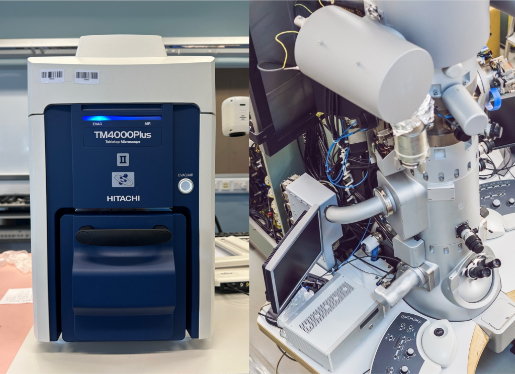
Yet, there was an unbreakable limit of optical microscopes no matter how sophisticated the optical microscope is - the wavelength of visible light. As the imaging source of optical microscopes is visible light, it is impossible to reveal the details being smaller than the wavelength of visible light. This limit was broken by Ernst Ruska, a German physicist who invented the first transmission electron microscope (TEM) in 1933. Different from optical microscopes, electron microscopes produce an enlarged image of objects using electron beams and magnetic lenses instead of visible light and optical lenses.
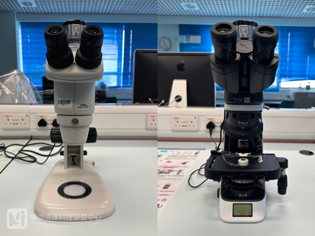
Today, there are various types of microscopes. Some of them are portable and are easy to handle. Microscopes have become one of the most important instruments for scientists. The development of microscopes will never stop.
Today, there are various types of microscopes. Some of them are portable and are easy to handle. Microscopes have become one of the most important instruments for scientists. The development of microscopes will never stop.
References:
American Physical Society (2004). Lens Crafters Circa 1590: Invention of the Microscope. Retrieved from https://www.aps.org/publications/apsnews/200403/history.cfm#:~:text=Lens%20Crafters%20Circa%201590%3A%20Invention,eyeglass%20maker%20named%20Zacharias%20Janssen
Davidson, M.W. (2010). Robert Hooke Physics, Architecture, Astronomy, Paleontology, Biology. Luminaries, 41(3), 180-182. Retrieved from https://academic.oup.com/labmed/article/41/3/180/2504959
Ford, B.J. (2007). Celebrating Leeuwenhoek’s 375th birthday – What could his microscope reveal? Retrieved from https://www.researchgate.net/publication/319388463_Celebrating_Leeuwenhoek’s_375th_birthday_-_What_could_his_microscopes_reveal
Science Museum (2019). The Microscope. Retrieved from https://www.sciencemuseum.org.uk/objects-and-stories/medicine/microscope
Source of photos
Figure 1. The Microscope Used by Robert Hooke
Credits: Micrographia: or some physiological descriptions of minute bodies made by magnifying glasses. With observations and inquiries thereupon / By R. Hooke. Wellcome Collection. Public Domain Mark
Figure 2. The Microscope Made by Anton van Leeuwenhoek (Replica)
Credits:Leeuwenhoek simple microscope (copy), Leyden, 1901-1930. Science Museum, London. Attribution 4.0 International (CC BY 4.0)
Figure 3. TEM (Right)
Credits: galitskaya (Canva creators), Transmission Electron Microscope in a Scientific Laboratory. Canva for Education
Author: Ms. CHIM Hoi Ying (Biology teacher)