Since the invention of the World’s first microscope, scientists have been observing and recording various objects with this tool. The advancement in imaging technology has changed the form of recording from manual drawing to photograph and video clip capturing, which enhances the accuracy and convenience for more extensive applications of the images.
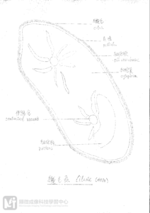
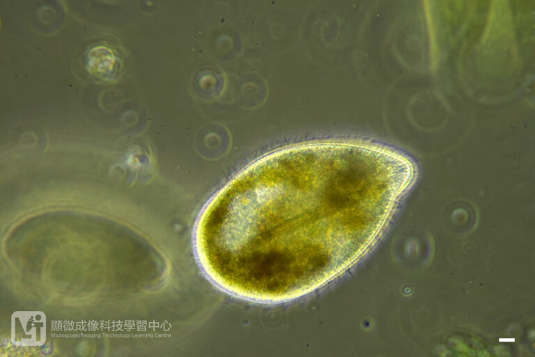
Adding imaging function to a microscope used for naked-eyed observation, so as to get accurate imaging record and measurement, is not as simple as casually connecting a camera to the eye piece. Yet, it requires sophisticated optical calculation and secured mounting.
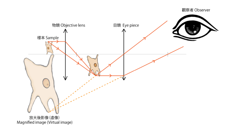
Referring to Figure 3, the magnified image produced by the compound microscope is a virtual image. Such virtual image cannot be projected onto any screens nor focused onto any camera sensors. If we want to observe a sample via a microscope and take photos at the same time, prisms are required to separate the light into two: one for observation and another for camera-recording. Moreover, groups of lens are also required to focus light rays onto the sensor thus forming real images, such method is called “Prime focus”; like we refract and focus the light rays from the eye piece with our eyes’ lens and retina during naked-eye observation. This additional set of prisms and lenses are installed in the “Trinocular tube”(Figure 4), thus this microscope is named “Trinocular microscope”(Figure 5).
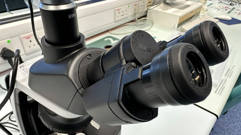
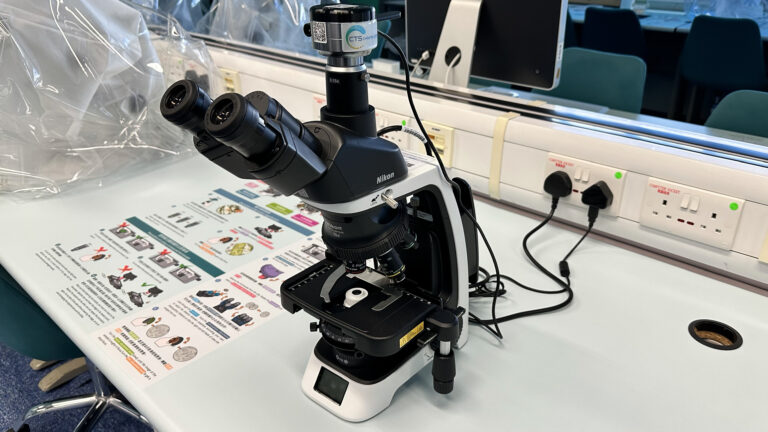
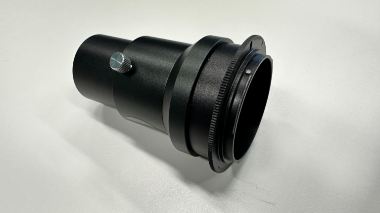
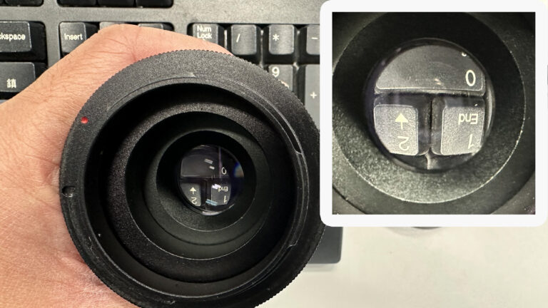
Once the focusing problem is sorted out, we could use cameras to record microscopic images. Nonetheless, when we connect DSLR or mirrorless cameras, which are possessed with larger sensors, to the microscope, we need to magnify the image in the microscope again until the image can be fully covered by the sensor. Addressing the market needs, some camera adapter tubes are manufactured with in-built lens group to meet the requirement of the higher resolution cameras (Fig. 6 & 7).
Author: Mr. CHAN Kam Kong (Biology teacher)


