I found a small insect on the floor in my living room. It is a tiny rice weevil and sized around 5mm, I cannot even observe its structure clearly with bare eyes. How does the rice weevil eat? What is the use of its long “nose”? I decided to bring it back to school and observe it in our Microscopic Imaging Technology Learning Centre.
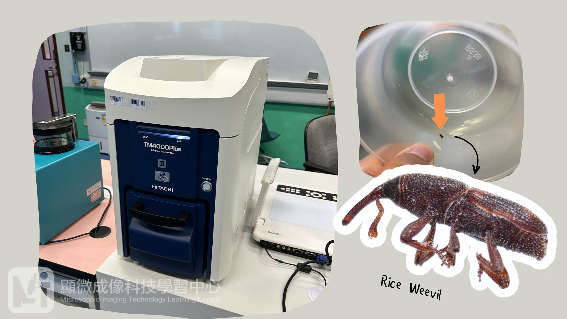
We can see rice weevil’s structure clearly under optical stereo microscope. Like other insects, rice weevil’ s body is divided into 3 parts, which are head, thorax and abdomen. Its head has many sensory organs like compound eyes and antennae. There are hair-like receptors on its antennae. Its body is darkish red in color and its abdomen is mostly covered by its elytron, which has orange patterns and small hairs on the surface.
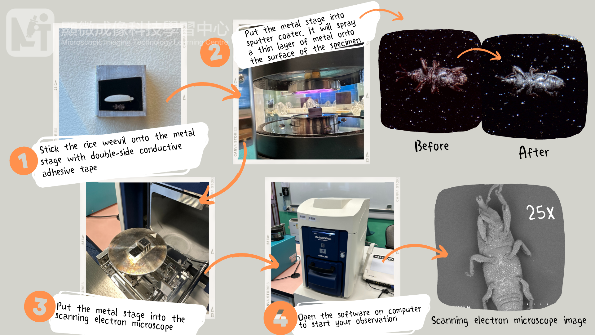
After observing the rice weevil under the optical stereo microscope, I still unable to figure out the long “nose” structure, so I decided to put it into the scanning electron microscope for closer observation. Using scanning electron microscope to observe insects is not as convenient as using optical stereo microscope, we need to prepare the sample beforehand with the following steps:
1) Take out the rice weevil from the fridge;
2) Stick it onto the metal stage with double-side conductive adhesive tape;
3) Put the metal stage in the sputter coater. It will spray a thin layer of metal on the surface of the sample which enable it to be conductive and create better images when using scanning electron microscope;
4) Put the metal stage into the scanning electron microscope and open on the software in computer to start your observation.
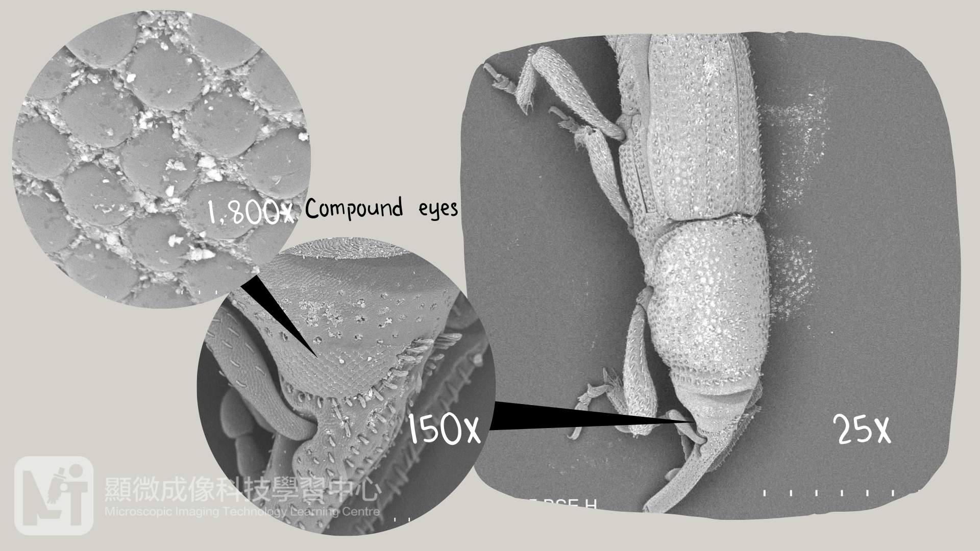
An optical microscope uses optical lenses to focalize the light source, and the light passes through the lens to magnify the image, while an scanning electron microscope uses magnetic lenses to refract electron beams, which scan the surface of the sample back and forth to generate signals and convert them into images. Since electron beam is not visible, the image of the scanning electron microscope is in monocolor. The magnification rate of the scanning electron microscope can reach to 10,000 times, which is much higher than that of the optical stereo microscope, which only can magnify 50 times. It allows us to observe the microstructure of the organisms.
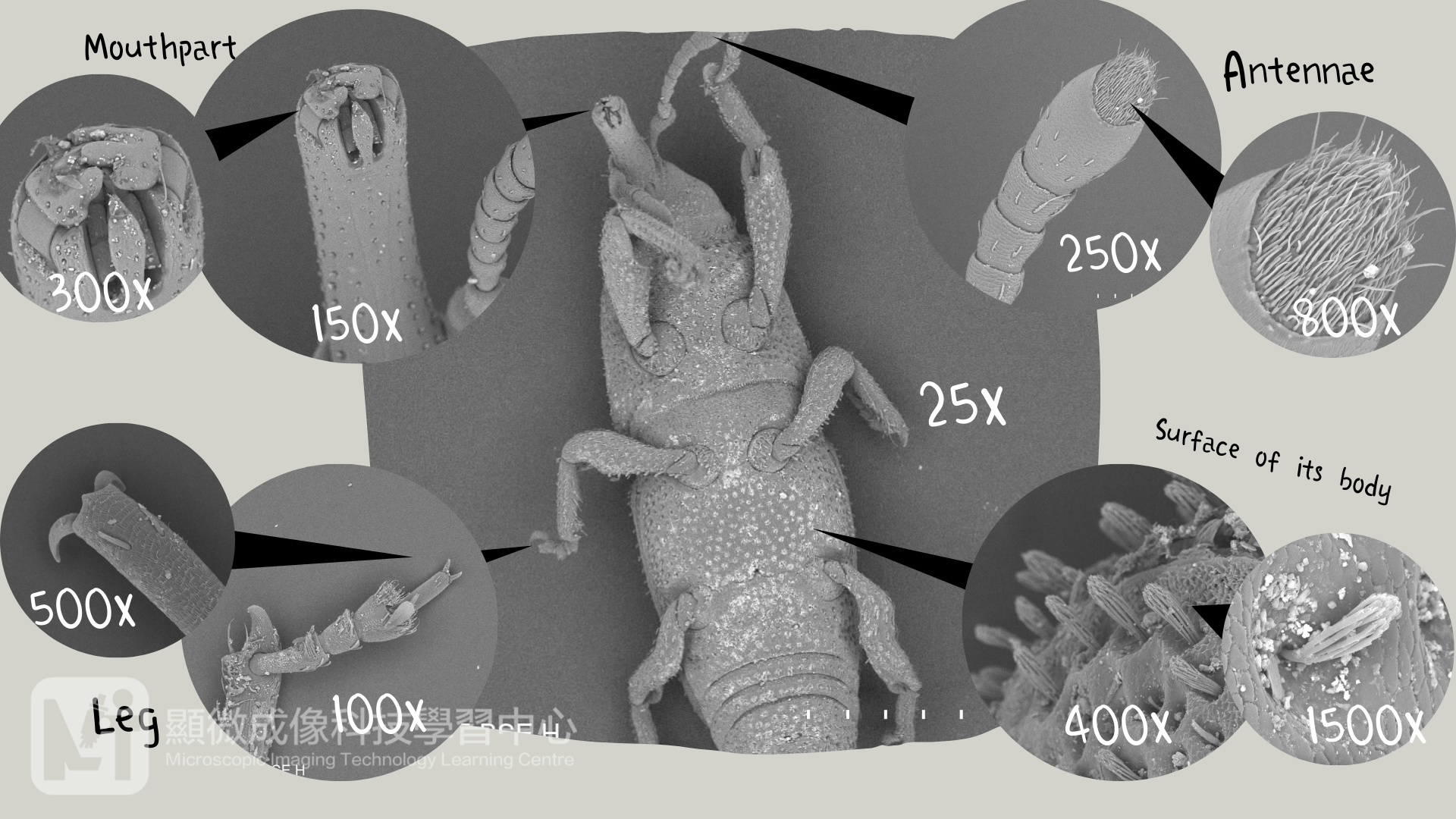
Scanning electron microscope is less flexible than optical stereo microscope, as we can not adjust the observation angle and direction freely. When I observe the back side of the rice weevil, I cannot see its mouthpart, so I stop the observation and take the sample out. Then flip it to the other side and repeat the sample preparation steps all over again. You can see different parts of the rice weevil in different magnification rate in Figure 4. If we have adequate samples, we might stick multiple number of insects onto the metal stage. Some of them with their back side facing up, some with their ventral aspect facing up, and some with their side facing up. We could conduct observation in different angles at the same time and save time for repeating the sample preparation.
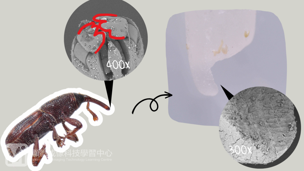
Now we know the answer! The long “nose” is actually rice weevil’s mouthpart. By observing with the scanning electron microscope, we can see there are two structures as if the alphabet “E”, it is the chewing mouthpart of the rice weevil. There are clear scratch marks on the rice grain, which are caused by its mouthpart.
Author: Ms. WONG Hoi Yan, Holly (Biology Teacher)


